New publication in Frontiers in Immunology
Photoconversion of fluorescent proteins enriches the toolbox to study cell migration in vivo. In a new paper Katja Jarick (née Ottmüller) describes how this procedure can be used to track alloreactive T cells migrating to graft-versus-host disease target tissues.
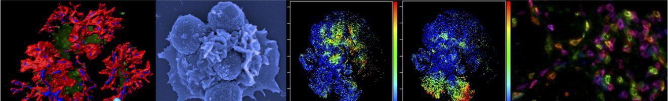
 Dendra2 is a fluorescent protein with fantastic features. In its natural state it provides green fluorescence. However, if cells expressing this fluorescent protein are exposed to light of a particular wavelength, their green color switches to red. In our paper by Jarick et al. we describe how this trick can be used to study the fate of effector T cells in mice. Dendra2 expressing cells can be photoconverted within tissues in real-time. Once photoconverted, they can be spatiotemporally tracked based on their unique color signature. Strikingly, even if T cells divided up to 4 times, we were still able to identify this photoconverted cells. Photoswitching of other fluorescent proteins has been reported before. However, Dendra2 proved optimal for T cell tracking due to high fluorescence quantum yields and low phototoxicity. Employing this technique now opens new avenues to study the fate of tumor infiltrating immune cell populations, cancer metastasis, migration patterns of allreactive T cells or the dynamics and plasticity of immune cell subsets in different scenarios such as infection, inflammation and immunotolerance. Our work has been generously supported by the IZKF Würzburg, the EFRE Program of the European Union and the DFG Transregio Research Network TRR221.
Reference: